Microbiology, the study of microorganisms such as bacteria, has been revolutionized by advances in imaging technology. One of the significant innovations in this field is the digital microscope. These powerful tools offer researchers and scientists a new way to explore the microscopic world of bacteria.
In this article, we’ll delve into the capabilities of digital microscopes, discuss the basics of purchasing one, and examine how they can be used to observe bacteria.
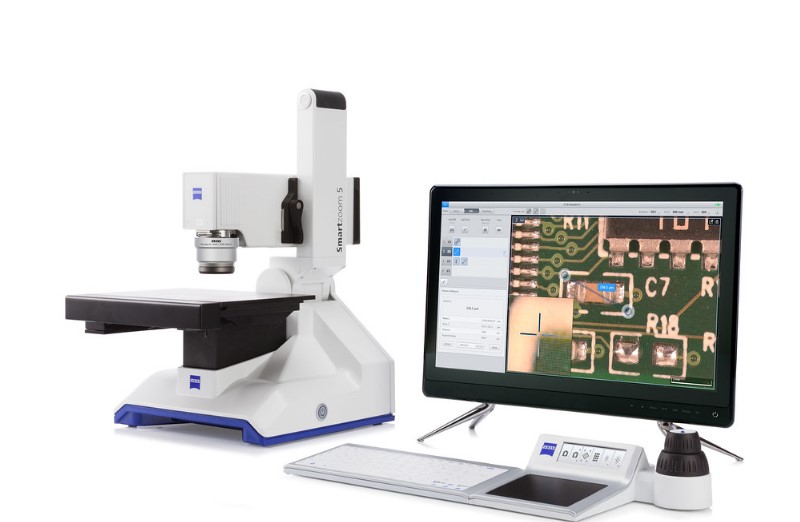
How Digital Microscopes Work?
Digital microscopes have gained popularity due to their ease of use and the convenience of digital image capture and sharing. Let’s explore how they work and why they are particularly well-suited for observing bacteria.
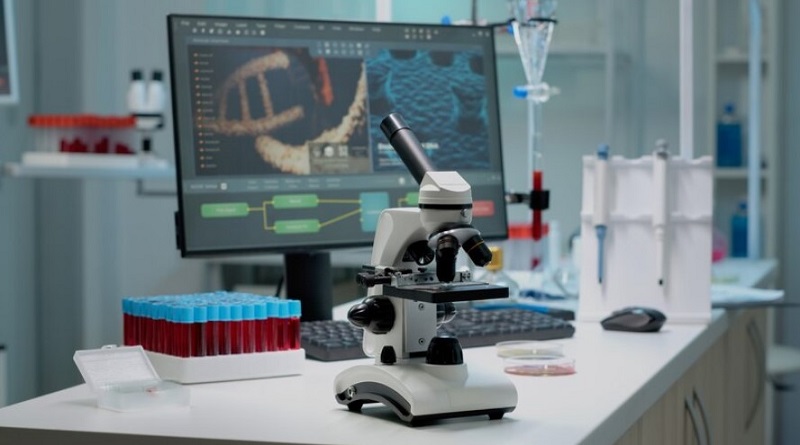
Digital microscopes combine traditional optical microscopy with digital imaging technology. They function by capturing images or videos of specimens through a built-in camera or external digital camera. These images can be displayed on a screen, saved for future reference, or shared with others.
Comparison to Traditional Optical Microscopes
Compared to traditional optical microscopes, digital microscopes offer several advantages. They eliminate the need for eyepieces, making them more user-friendly and comfortable for extended use. Additionally, digital microscopes allow for easy image capture and analysis, making them an excellent choice for research and educational purposes.
Advantages of Digital Microscopes in Observing Bacteria
Digital microscopes excel in observing bacteria for several reasons:
- Digital Imaging: The primary advantage of digital microscopes in studying bacteria is their ability to capture high-quality digital images and videos. This feature is invaluable for researchers as it enables them to document their findings for future reference and share them with colleagues or students effortlessly. This digital documentation can serve as a valuable resource for educational purposes and collaborative research.
- Live Imaging: In addition to static imaging, some digital microscopes offer live imaging capabilities. This feature allows scientists to observe dynamic processes within bacterial cells in real time. Whether it’s the movement of flagella or the replication of bacterial colonies, live imaging provides critical insights into the behavior and life cycles of bacteria. Researchers can gain a deeper understanding of bacterial processes by observing them as they happen.
- Measurement and Analysis: Many digital microscopes are accompanied by sophisticated imaging software. This software not only aids in capturing images but also allows for precise measurement and in-depth analysis of bacterial specimens. Researchers can quantify various parameters, such as bacterial size, shape, and motility. This quantitative approach is especially valuable for conducting rigorous scientific experiments and studies in microbiology.
- Data Integration: Digital microscopes facilitate the integration of imaging data into broader research workflows. Researchers can easily link microscope-generated data with other analytical tools and software, streamlining data processing and analysis. This interconnected approach enhances the efficiency and accuracy of bacterial research, contributing to a deeper understanding of these microorganisms.
- Remote Collaboration: The digital nature of these microscopes allows for remote collaboration. Researchers and scientists from different locations can simultaneously view and discuss bacterial observations in real-time. This feature is particularly beneficial in today’s interconnected scientific community, as it fosters collaboration and knowledge sharing among experts in the field.
Bacteria Size and Characteristics
Before we dive into the practical aspects of observing bacteria with digital microscopes, it’s essential to understand the characteristics of these tiny microorganisms. Bacteria are single-celled microorganisms that are found nearly everywhere on Earth. They play essential roles in various ecosystems, and some species can be harmful pathogens. Understanding their structure and behavior is crucial for various scientific fields.
Bacteria are incredibly small, typically ranging from 0.5 to 5 micrometers in size. To put this in perspective, thousands of bacterial cells could fit within the width of a human hair. Observing bacteria at such a scale requires a microscope with high magnification capabilities. Bacteria come in various shapes and sizes. Some common morphological features include:
- Cocci: Spherical bacteria
- Bacilli: Rod-shaped bacteria
- Spirilla: Spiral-shaped bacteria
Visualizing Bacteria with Digital Microscopes
Visualizing bacteria with digital microscopes involves several key steps to achieve clear and detailed observations. Digital microscopes provide a wide range of magnification options, allowing scientists to precisely zoom in on bacterial specimens. They excel in providing excellent resolution, making it possible to visualize intricate details within bacterial cells, such as cell walls, flagella, or cilia.
However, achieving such clarity requires careful sample preparation, which includes fixing and staining the bacteria. Staining is essential to enhance contrast and visibility, highlighting specific structures within bacterial cells. Various staining methods, including well-known techniques like Gram staining, enable researchers to differentiate between different types of bacteria based on the color or intensity of the stain, making the study of bacteria more effective and informative.
Basics to Buy a Microscope
When buying a microscope, start by understanding your specific needs and budget. Digital microscopes come in various models, suited for different purposes, from educational to advanced scientific research. You’ll find two primary microscope categories: optical and digital. Optical microscopes use lenses for magnification and are great for observing transparent or stained specimens.
In contrast, digital microscopes are equipped with digital cameras and screens, facilitating image capture and sharing. For bacterial observation, focus on high magnification, resolution for clear details, proper lighting (brightfield, phase contrast, or darkfield), and digital imaging capabilities, enabling you to document and share your findings effectively. You can read the complete details here on this page.
Conclusion
Digital microscopes have opened up new possibilities in the field of microbiology, allowing researchers to observe and document bacterial cells with unprecedented ease and precision. While they have their limitations, digital microscopes continue to play a crucial role in expanding our understanding of bacteria, driving advancements in medical diagnostics, and supporting environmental studies. As technology advances, the future of bacterial observation with digital microscopes holds even more promise, offering exciting opportunities for scientific discovery and innovation.